标签打印机打印错位(儿童锁骨骨折,保守治疗,骨折错位,家属不满意,咋办?)
Posted
篇首语:时穷节乃现,一一垂丹青。本文由小常识网(cha138.com)小编为大家整理,主要介绍了标签打印机打印错位(儿童锁骨骨折,保守治疗,骨折错位,家属不满意,咋办?)相关的知识,希望对你有一定的参考价值。
标签打印机打印错位(儿童锁骨骨折,保守治疗,骨折错位,家属不满意,咋办?)
儿童锁骨骨折,保守治疗,骨折错位,家属不满意,咋办?产伤以及婴幼儿锁骨骨折如何治疗?
在临床工作中,产伤,以及婴幼儿骨折,很多骨科医生并不知道如何处理。看到有移位的骨折,骨科医生就建议手术,甚或直接开刀手术,家属担心骨折对位不齐,会影响孩子的发育而左右医生的决策。本文结合儿童锁骨骨折临床实例在小儿骨科医生之间的讨论以及对Rang's Children's Fractures 4th Edition中关于婴幼儿锁骨骨折治疗的论述,希望对于骨科医生,妇产科医生以及患儿家属有一定的帮助作用。
中华骨科网-儿童骨科1群 微信群上的聊天记录
—————2022-08-31 —————
马扩助 临沂市第三人民医院 09:40
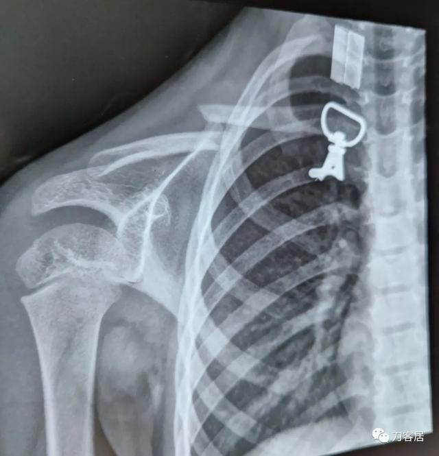
大家好,儿童锁骨骨折,保守治疗,八字绷带,家属不满意,大家都是怎么处理的。谢谢。

徐丰~开封市儿童医院 09:47
几岁?多长时间了?
马扩助 临沂市第三人民医院 09:51
5岁,一周
马扩助 临沂市第三人民医院 09:52
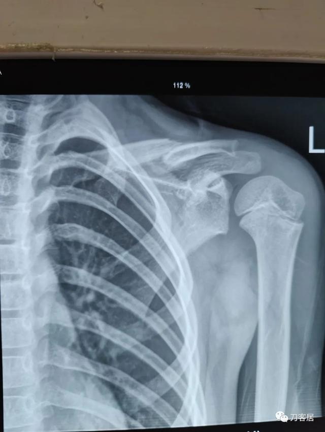
这是今天第二个锁骨保守,7岁,同样8字固定,一个月了,家长不满意。
两个都是女孩
周晓康河北省儿童医院 09:53
只要耐心多解释清楚,看看半年后的片子,家属就会容易理解满意。 如果只是看近期的片子,外行是不会满意的
洪攀 武汉协和骨科 09:57
不满意 解释一下,毕竟没有手术指证,佛度有缘人,不信任你,也就不用太多废话。
马扩助 临沂市第三人民医院 09:57
[握手][握手]@河北省儿童医院,周晓康
@洪攀 武汉协和小儿骨科 谢谢
山大二院柳晓军 10:00
后期会塑型很好的,可以让家长看些治疗过的片子,减少焦虑。
燕华—华新儿骨 10:10
为了避免家属不满意,可以考虑伤后一个月不要拍片子。
ZRH 10:19
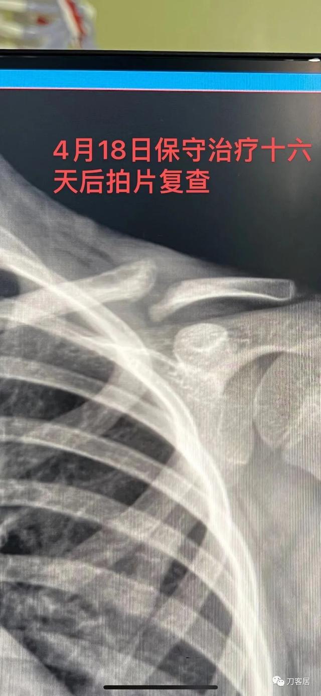
大家都很敬业苦口婆心,奈何家属可能不理解,我提供个反面教材给看看手术失败病例,也让家属感受一下。


马扩助 临沂市第三人民医院 10:38
@燕华—华新儿骨 谢谢
吕洪海 10:38
好病例,值得收藏
马扩助 临沂市第三人民医院 10:38
@武汉协和医院骨科迮仁浩 谢谢
吕洪海 10:40
医生的治疗方案不能被家属左右,按照原则办事
陈建松浙大儿院骨科 10:40
请问这个病例,你最后怎么处理了?
马扩助 临沂市第三人民医院 10:41
这种保守治疗,儿骨医生和骨科医生一般认为是很好的,就怕不良医生或外行随便点评。
ZRH 10:41
网络问诊的病例,没有后续,但觉得孩子很无辜!
洪攀 武汉协和骨科 10:42
@武汉协和医院骨科迮仁浩 这个是几岁的小朋友呀
ZRH 10:46

徐丰~开封市儿童医院 10:47
@临沂三院骨科马扩助 对于这个患儿,绷带可以经常紧一紧,别太松。
山大二院柳晓军 10:54
还有两点,一去枕平卧可以使得锁骨保持平整,二要固定上臂,这样有效固定断端移位。
Tony 10:54
为啥要急着拔针?针留在皮外?
ZRH 10:56
网络问诊的病人,我也不知道那个医院为啥要这么快拔针[难过],应该是术后8周,见骨痂生长,皮外针尾也有刺激,就考虑拔针了。
Tony 11:02
目前这个病人从片子上看确实得要植骨了,骨不连很明显了,髓腔断端看上去也很圆滑,似乎已经闭合了[捂脸]。
陈建松浙大儿院骨科 11:04
植骨的话,怎么固定,用钢板吗?
Tony 11:05
我觉得还是用克氏针可靠,骨膜剥离少,血运破坏少,有利愈合。
ZRH 11:11
植骨,打通髓腔,个人可能更倾向稳定性更好的接骨板,骨折端微动不稳定可能还是不愈合。处理类似先天性锁骨假关节。
洪攀 武汉协和骨科 11:14
@武汉协和医院骨科迮仁浩 应该追求稳定,用锁定板固定,取髂骨,类似先天性锁骨假关节,上肢和下肢骨折不愈合应该治疗理念存在一定的差异。
Tony 11:23
追求足够的稳定,可以考虑锁定钢板,用了锁定板,打通髓腔后,是否还需要植骨?[捂脸]
洮河刀客 11:27
@临沂三院骨科马扩助 有一张图没发出来?看不到
马扩助 临沂市第三人民医院 11:29
马老师,前面两个片子是一个5岁女孩的,后面那个片子是一个7岁女孩的,只有一张。
洪攀 武汉协和骨科 11:29
@刘喜平-湖南省儿童医院骨科 不容有失,植骨稳妥。
马扩助 临沂市第三人民医院 11:30
找到了,是这个。

Tony 11:30
[憨笑][强]@洪攀 武汉协和小儿骨科 现在的医生最担心的就是怕不稳妥,怕惹麻烦[微笑]。
洪攀 武汉协和骨科 11:31
佛度有缘人,说清楚,其他的,随他去。
吕洪海 11:32
儿童锁骨骨折罕见不愈合,这个孩子锁骨骨折不愈合,与手术广泛剥离,骨膜损伤有关系。
洪攀 武汉协和骨科 11:33
保守治疗几乎没有不愈合的。
吕洪海 11:34
总有无知无畏者,拿孩子健康练手艺,可悲可怕!
Tony 11:34
打根克氏针需要广泛剥离骨膜吗?[捂脸] 术者那样的打法估计闭合都能打上。
小米虫 11:37
如果是切开复位的话是有可能剥离的。
Tony 11:38
嗯嗯,是的
ZRH 11:40
应该是切开复位的,克氏针的尖都去掉了,闭合复位就很难穿进去,只是不知道剥离了多少。
Tony 11:42
也可能是先打的外端,从髓腔破骨皮质出,再倒打入内侧端,所以就没有针尖了[捂脸]。
吕洪海 11:45
看术后片子,术者可能是切开手术。
Tony 11:48
不愈合的话,估计是切开复位的[微笑],我目前还没有见到过闭合复位不愈合的病人。
洮河刀客 12:00
@临沂三院骨科马扩助 好的。谢谢你了
赵黎英华儿童骨科医生集团 12:02
同意
下面是Rang's Children's Fractures 4th Edition中关于婴幼儿锁骨骨折治疗的论述。
Rang's小儿骨折第四版中婴儿锁骨骨折的治疗方案。分别对锁骨中段骨折、内外侧(远端)骨折进行了说明。特别将婴儿(Infancy)锁骨骨折单独一段说明。
Infancy
婴幼儿锁骨骨折
Clavicle fractures are one of the most common injuries sustained during childbirth; children of large birth weight (greater than 4,000 g) and those with shoulder dystocia are at the highest risk. Infants who sustain a clavicle fracture may also sustain a brachial plexus injury because of nerve stretch (Erb palsy). The neonate with a clavicle fracture may present with an asymmetric Moro reflex or the appearance of a flail upper extremity.
锁骨骨折是分娩过程中最常见的损伤之一;体重较大(大于4000克)新生儿和肩部难产新生儿发生锁骨骨折的风险最高。锁骨骨折的新生儿也可因神经牵拉而导致臂丛神经损伤(Erb麻痹)。锁骨骨折的新生儿可能出现不对称莫罗反射(新生儿的拥抱反射)或连枷上肢外观。
Differentiating a neurologic injury from a clavicle fracture during the first few weeks of life can be extremely difficult, and the child may have both. Xray or ultrasound can diagnose the fracture, but clear neurologic assessment of the upper extremity may not be possible until the fracture has healed.
出生后的最初几周内,鉴别臂丛神经损伤和锁骨骨折可能非常困难,而且新生儿可能同时存在这两种情况。X线或超声波可以诊断骨折,但在骨折愈合之前,无法对上肢神经进行明确评估。


Ernst Moro (1874-1951):Dr. Moro was an Austrian pediatrician who described a defensive infantile reflex normally present in all infants/newborns up to 3 or 4 months of age. When the infant feels as if they are falling, they immediately abduct the arms, and then draw their arms across their chest in an embracing manner. An asymmetric Moro reflex may be secondary to neurologic injury or fracture.
恩斯特·莫罗(1874-1951):莫罗医生是一位奥地利儿科医生,他描述了所有3个月或4个月大的婴儿/新生儿通常都会出现的婴儿防御反射。当婴儿感觉自己好像要跌落时,他们会立即外展手臂在胸前呈拥抱状。不对称莫罗反射可能继发于神经损伤或骨折。
Some children are born with a congenital pseudoarthrosis of the clavicle (Fig.6-3). This can easily be confused with a clavicle fracture and has instigated unnecessary child abuse work-ups. The painless swelling over the midshaft of the clavicle is often noted in infancy but may go undetected for years. The xray will show a smooth intact cortex at the site of pseudoarthrosis and not the jagged edges of acute fracture. A fracture will have abundant callus within a few weeks, and no callus will develop if it is a pseudoarthrosis.
一些新生儿出生时即存在先天性锁骨假关节(图6-3),易误诊为锁骨骨折,并引发不必要的对是否存在虐待儿童行为进行调查。锁骨中段的无痛肿块通常在婴儿期出现,但可存在数年而未能发现。X线片显示假关节部位骨皮质光滑完整,而不是急性骨折的锯齿状边缘。骨折在几周内会有大量的骨痂,而假关节不会形成骨痂。

Figure 6-3 Congenital pseudoarthrosis of the clavicle can easily be confused with a fracture. 图6-3先天性锁骨假关节易误诊为锁骨骨折。
The majority of children with clavicle congenital pseudoarthrosis do well with no treatment. However, if the patient does become symptomatic or the parents are unhappy with the appearance, resecting the pseudoarthrosis, bone grafting, and plating provide predictable union.
大多数小儿锁骨先天性假关节无需特殊治疗,对其正常发育和活动没有多大影响。然而,如果患儿有症状或父母对其外观不满意,可选择切除假关节、植骨和钢板固定,一般都会取得良好的愈合。
Children and Adolescents
儿童及青少年锁骨骨折
Most clavicle fractures occur as a result of a fall directly on the shoulder with the arm at the side, but less commonly a fracture may occur as a result of a direct blow or a fall on an outstretched hand. Participation in contact sports such as football, rugby, wrestling, and hockey are responsible for the largest percentage of clavicle fractures in adolescence, but with our nation’s increased interest in extreme sports such as BMX, motocross, and mixed martial arts (MMA), we are seeing higher-energy fractures more frequently.
大多数锁骨骨折是由于摔倒时手臂在身侧,肩部直接着地所致,但也有一些不太常见的骨折可能系由直接打击或上肢外展时着地所致。青少年锁骨骨折最常见的原因是参加碰撞性运动,如足球、橄榄球、摔跤和曲棍球,但随着极限运动,如越野自行车(BMX)、越野摩托车和综合格斗(MMA)的普及,可见更多高能量损伤的锁骨骨折。
The examination of a child with a clavicle fracture is relatively straight forward given the superficial nature of the bone. Typically, the patient will present with the arm being held in an adducted position close to the body with the opposite hand supporting the injured extremity. The skin should be inspected for an open fracture or significant tenting (which has rarely been reported to erode through the skin). Typically, the clinical deformity, ecchymosis, swelling (Fig. 6-4), and point tenderness lead the physician to the diagnosis.
鉴于锁骨位置浅表,儿童锁骨骨折的检查相对直接。通常,患者会将手臂贴近身体置内收位,健侧手扶患侧上肢。应检查皮肤以判断有无开放性骨折或明显的局部隆起(很少有刺破皮肤的报道)。局部畸形、瘀斑、肿胀(图6-4)和压痛即可诊断锁骨骨折。

Figure 6-4 The clavicle is subcutaneous, making deformity noticeable. This patient has a healing right clavicle fracture. Patients need to be told about the size of callus that will appear (and later resorb). 图6-4 锁骨位皮下,因此畸形明显。此患者右侧锁骨骨折正在愈合。需告知患者将出现的骨痂大小(以及随后的再吸收)。
Limb threatening concerns associated with clavicle fractures and dislocations that need to be identified immediately include vascular injury (subclavian vessels), neurologic injury (brachial plexus), and injury to the mediastinal structures (esophagus, trachea, pleura, and lung) by angulated or displaced fragments.
对于锁骨骨折脱位成角或粉碎移位导致的并发症需要进行检查,包括血管损伤(锁骨下血管)、神经损伤(臂丛)和纵隔结构损伤(食管、气管、胸膜和肺)。
CLASSIFICATION
锁骨骨折分型
Fractures can be complete, incomplete but angulated, or plastically deformed (Fig. 6-5). The very thick layer of periosteum surrounding the pediatric clavicle tends to maintain the alignment of the fracture, which typically leads to early union in infants and children. As children become teenagers, the periosteum no longer acts as a strong supporting structure leading to greater fracture displacement and a higher risk of non-union.
骨折可分为不全骨折、完全骨折、不全成角骨折,青枝骨折畸形(图6-5)。小儿锁骨周围厚骨膜层保持骨折不易移位,使婴儿和儿童易早期愈合。随着儿童成为青少年,骨膜变薄,不再是一个强有力的稳定结构,骨折容易移位,骨折不愈合风险也较高。
儿童锁骨骨折的分型

锁骨骨折的基本类型
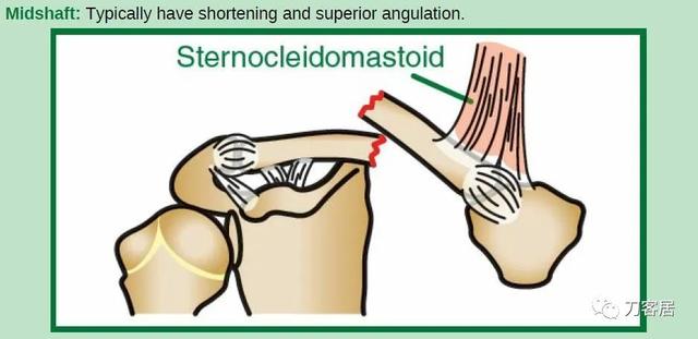
中段骨折典型表现:短缩及向上成角畸形。
Lateral: Further subdivided by Dameron and Rockwood.
Note: The epiphysis and periosteum typically remain in place and the shaft displaces.
外侧骨折:由Dameron和Rockwood进一步分型。
注:骨骺和骨膜通常保持原位,骨干移位。

I: No significant displacement,I型:无明显移位

II: Mild displacement(<25%),II型:轻度移位(<25%)

III: Superior displacement(25%-100%),III型:向上移位(25%-100%)

IV: Posterior displacement,IV型:向后移位

V: Superior displacement (>100%),V型:向上移位(>100%)
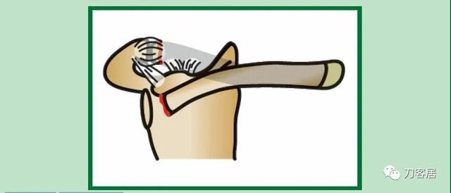
VI: Inferior displacement,VI型:向下移位
Medial: Subdivision of medial clavicle fractures. The description of the fracture can be based on displacement of the shaft—anterior, posterior, of inferior.
内侧:锁骨内侧骨折进一步分型。骨折分型基于骨干向前、向后和向下移位。

A: Physeal fracture,A: 骺骨折

B: Sternoclavicular dislocation (rare),B: 胸锁关节脱位(少见)

C: Medial shaft fracture,C: 内侧骨干骨折
The basic types of fracture include medial, lateral, and midshaft fractures. Medial and lateral fractures have been further subdivided based on location of the fracture and displacement of the shaft (see “Classification of Pediatric Clavicle Fractures” for an overview of the sub-classifications).
骨折的基本分型包括内侧、外侧和中段骨折。根据骨折位置和骨干移位方向,进一步分为内侧和外侧骨折(有关亚型分型,请参阅“儿童锁骨骨折的分型”)。
TREATMENT—MIDSHAFT FRACTURES
治疗-中段骨折
The periosteum is much thicker, stronger, and less readily torn in a child than in an adult and continuity of the periosteum determines whether or not a fracture displaces. When displacement occurs, the intact hinge of periosteum can help or hinder reduction.
与成人相比,儿童的骨膜更厚、更坚固、更不易撕裂,骨膜的连续性决定了骨折是否移位。发生移位时,完整的骨膜铰链有助于复位,也可阻碍复位。
Infant
Infant clavicular fractures can be treated by pinning the shirtsleeve to the shirt (Fig. 6-6) or loosely wrapping the arm to the body with an elastic bandage for 2-3 weeks. This treatment provides some immobilization and pain relief and reminds people not to pick the baby up by the arm. Infantile fractures tend to heal well regardless of treatment. The associated injuries including brachial plexus palsy require more focused attention; however, these are difficult to evaluate until the fracture heals and motion can be better assessed.
婴幼儿锁骨骨折
婴幼儿锁骨骨折可以通过将患侧衣服袖子固定在衣服上(图 6-6)或用弹性绷带将手臂松散包裹固定于身体上2-3 周进行治疗。此固定可缓解疼痛,并提醒抱起患儿时不要提患儿的患肢。不管治疗与否,婴幼儿骨折往往愈合良好。(如并发臂丛神经损伤,则需特别注意;但是在骨折时很难进行评估,需待骨折愈合患侧上肢可以自主活动后,方可进行评估。)

Figure 6-6 An infant with a clavicle fracture can be treated by pinning the sleeve (of the injured side) to the body of the garment. A second option: wrap the limb to the trunk gently with an ACE bandage.图6-6.婴幼儿锁骨骨折可以通过将患侧衣服袖子固定在衣服上或用弹性绷带将手臂松散包裹固定于身体上治疗
Children and Adolescents
儿童和青少年锁骨骨折
Current trends in orthopedic care suggest that treatment selection for midshaft clavicle fractures has become more controversial. Historically, indications for surgical fixation were relatively limited including open fractures and severely displaced fractures with significant skin tenting or neurovascular compromise.
儿童和青少年锁骨中段骨折的治疗选择存在争议。从历史上看,手术固定的适应证相对有限,包括开放性骨折和严重移位的骨折,伴有明显的皮肤隆起或神经血管损伤。
With the publication of several randomized controlled trials in adult populations showing faster healing rates, lower non-union rates, and better functional outcomes with surgical intervention, many surgeons have been applying these “adult principles” to adolescent and pediatric patients. Currently, the literature is unclear as to which adolescent clavicle fractures should be fixed. As an institution, we trend toward non-operative treatment for the vast majority of clavicle fractures.
有几项针对成人人群的随机对照研究显示,手术可促进愈合,提高治愈率、降低不愈合率,可获得更好的功能结果。许多外科医生将这些“成人原则”应用于青少年和儿童患者。目前,关于青少年锁骨骨折是否需要手术固定的文献尚不清楚。我们对绝大多数锁骨骨折进行非手术治疗。
Nearly all minimally displaced clavicle fractures can be treated with a sling or a figure-of-8 brace (Table 6-2). A theoretical advantage of the figure-of-8 is that it potentially pulls the shoulders back minimizing fracture fragment overlap. A practical advantage is that it frees the extremity making simple daily tasks such as computing easier. Practical advantages of the sling, on the other hand, include its ease of use, ubiquitous availability, and cost effectiveness.
几乎所有轻微移位的锁骨骨折都可使用吊带或8字形绷带固定进行治疗(表 6-2)。8字形绷带可将肩部向后拉,从而最大限度地减少骨折片段的重叠,其优势是解放了肢体,可以完成日常简单的任务,比如写作业和使用电脑。而悬吊带固定的优势是方便获得、操作简单、实用、廉价。
Many clinical trials failed to show significant outcome differences between slings and figure-of-8 braces. As a result, there are regional preferences for one or the other.
许多临床试验并未证明前臂吊带和 8 字形绷带之间存在显著差异。因此,不同的区域的医生会有不同的选择。
“While surgical fixation may return athletes with displaced fracture to sport faster, we do not believe that the four to six weeks gained justifies the vastly greater treatment costs, risks of surgery, and likely need for implant removal (that will take them out of sports again) for most amateur athletes”
“虽然手术固定可能会使骨折移位的运动员更快地恢复运动,但我们认为,对于大多数业余运动员来说,4到6周的保守治疗,与手术治疗需要更多费用、承担更多更大的手术风险以及可能再次手术去除植入物(这将使他们再次退出运动场)相比,更值得一些。”
Table 6-2 Classic Dilemma: Sling versus Figure-of-8 Brace 表6-2典型困境:吊带还是8字绷带固定 | ||
Advantages优点 | Disadvantages缺点 | |
Sling,吊带固定: 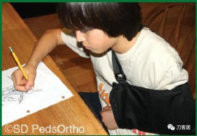 | Very inexpensive 特便宜 Easy to put on 容易佩戴 No pressure over fracture 骨折处无压迫 A few sizes fit all 几种尺码适合所有人 | No ability to pull fracture to length 不能恢复骨折长度 Hand is not free 手不自由 |
Figure-of-8, 8字绷带固定  | Can hold fracture better reduced (in theory) (理论上)可维持骨折复位 Hands free for activities 手自由活动 | Harder to put on 不容易佩戴 Focal pressure over fracture site 骨折局部有压迫 Need to keep multiple sizes in stock 需要库存多种尺寸 |
When a non-operative approach is utilized, the fracture is protected for 4-6 weeks, with contact sports avoided for another 6 weeks. As in most simple injuries, half the treatment consists of educating the parents about the normal course. An unsightly lump may appear with fracture healing (callus) and will potentially persist for a year while remodeling progresses (we tell parents that the lump may be the size of a walnut or an egg—Fig.6-7).
当采用非手术方法时,骨折固定 4-6 周,另外6周内避免碰撞运动。与大多数简单的损伤一样,一半的治疗包括对患儿父母进行正常骨折愈合过程的教育。在骨折愈合(骨痂)时可能会出现难看的包块,并且在重塑过程中可能会持续一年(我们应该告诉患儿父母,包块可能有核桃或鸡蛋那么大(图 6-7)。
Figure 6-7 Significantly overlapped midshaft clavicle fracture in a teenager. We warn patients that the resulting callus may be the size of a walnut (or even an egg in a teenager). With time, most fractures remodel nicely.图 6-7 青少年明显重叠的中段锁骨骨折。我们应该告知患者,由此产生的骨痂组织可能有核桃大小(有的可能有鸡蛋大小)。随着时间的推移,大多数骨折都能很好地重塑。
Although x-rays of a fracture healing in bayonet opposition may frighten the parents, studies have shown that a significant amount of angulation and overlap can be accepted. Once the fracture is non-tender and there is radiographic healing, the patient may slowly return to sports. Final x-rays are usually obtained at 4-6 weeks after injury; if there are concerns of a developing non-union, longer follow-up becomes necessary.
虽然X线片上错位成角骨折愈合可能会吓到患儿父母,但研究表明,较大的成角和重叠可以接受。一旦骨折无痛,且X线片显示有骨愈合,患者可逐渐恢复运动。通常在伤后4-6周行最后一次X线检查;如果存在不愈合可能,则需要更长时间的随访。
Surgical Reduction?
There are four primary concerns that drive patients and their families toward surgery:
1. Concern that a malunion will lead to functional deficits
2. Concern about developing a non-union
3. The cosmetic concerns discussed previously
4. The concern that a non-operative approach will take longer to heal
四个方面的顾虑促使患者及其家属接受手术:(译者注:这些同时往往成了忽悠或诱导患儿家长接受手术的理由)
1.担心畸形愈合导致功能缺陷;
2.担心骨折不愈合;
3.不美观;
4.担心非手术方法需要更长的时间才能愈合。
Concerns over a symptomatic malunion are possibly the strongest indication for surgery, but still not well validated in the pediatric or adolescent literature. Various criteria for surgery have been proposed including complete displacement, greater than 2 cm of shortening, and comminuted fracture patterns. Our institution, as well as Boston Children’s Hospital, have published studies showing that patients treated non-operatively (even with significant displacement and shortening) have no significant functional deficits and are able to return to high levels of overhead sport. Taking into consideration that the rare established symptomatic malunion can still be managed with late surgery with good outcomes, nearly all of these fractures can be treated without surgery.
担心会发生症状性畸形愈合可能是手术的最强指征,但在小儿以及青少年骨折治疗文献中并未得到很好的验证。现有的手术适应证包括完全移位、大于2cm的短缩和粉碎性骨折。但我们机构以及波士顿儿童医院发表的研究表明,非手术治疗的患者(即使有明显的移位和短缩)并没有明显的功能缺陷,并且能够恢复高水平的高于头颅平面的运动。考虑到罕见的症状性畸形愈合可通过晚期手术获得良好的结果,几乎所有这些骨折都可以进行非手术治疗。

Figure 6-8 Although rare, clavicle nonunions can occur in children.
虽然罕见,但小儿锁骨骨折确实有骨不连发生的可能。
Although non-unions have been reported in as many as 15% of completely displaced clavicle fractures in the adult population, they remain an extremely rare complication in pediatric patients with less than a dozen cases having been reported in the literature (Fig. 6-8). Over the last 10 years, our institution has treated hundreds of midshaft clavicle fractures and we have only observed three non-unions all of which were successfully managed with local bone graft and plate fixation. We, therefore, do not believe that nonunion concerns in adolescent patients, even with displaced fracture patterns, justify acute surgery.
尽管成人完全移位的锁骨骨折存在15%的不愈合,但儿童患者完全移位的锁骨骨折不愈合极为罕见,文献报道的病例不到十几例(图6-8)。过去10年里,本单位治疗的数百例锁骨中段骨折,只有3例骨折不愈合,所有这些骨折不愈合均通过局部植骨和钢板固定成功治愈。因此,我们不认为青少年患者的锁骨骨折骨不连是个问题,即使骨折移位,也不能作为急性手术的理由。
Although surgical fixation may return athletes with displaced fracture to sport faster, we do not believe that the 4-6 weeks gained justifies the vastly greater treatment costs, risks of surgery, and likely need for implant removal (that will take them out of sports again) for most amateur athletes.
虽然固定手术可使骨折移位的运动员更快地恢复运动,但我们认为,对于大多数业余运动员来说,保守治疗4-6周的时间,与手术相关的治疗费用、手术风险以及可能需要内植物去除手术(这将使患者再次退出运动)相比,更划算一些。
For the rare midshaft clavicle fracture requiring surgery, plate fixation can be used for all fracture patterns. The rigid construct enables early mobilization and a rapid return to sports. Some centers are now using intramedullary stabilization with elastic nails for non-comminuted fracture patterns (Fig. 6-9); this minimizes the scar that results from open treatment. To date, no study has compared the results of plate fixation to intramedullary fixation in the pediatric population.
对于罕见的需要手术的锁骨中段骨折,钢板固定可用于所有骨折类型。刚性结构使早期活动和快速恢复运动成为可能。一些中心现在使用弹性钉进行髓内固定治疗非粉碎性骨折(图6-9);这可以最大限度地减少开放治疗造成的疤痕。迄今为止,尚无小儿锁骨严重移位骨折行钢板内固定和髓内固定的比较性研究。

Figure 6-9 Intramedullary devices offer an alternative to plate fixation for the rare surgical fracture. (Image courtesy of Chris Souder, MD.)罕见锁骨骨折手术,使用弹性锁骨髓内钉代替钢板内固定。
TREATMENT—LATERAL FRACTURES
Dameron and Rockwood suggest that Type I, II, and III distal clavicle fractures will heal and remodel without intervention. Reduction and fixation of these lateral-sided injuries is only necessary for Types IV, V, and VI that have a severe and fixed deformity. Distal clavicle injuries in pediatric patients are usually transphyseal fractures and not true AC separations (as seen in adults). The intact periosteum allows children to heal and remodel with few complications without operative intervention.
外侧骨折的治疗
Dameron和Rockwood建议I、II和III型锁骨外侧骨折无需干预即可愈合和重塑。仅对具有严重和固定畸形的IV、V和VI型才需要干预。小儿患者的远端锁骨损伤通常是骨骺骨折,而不是真正的AC分离(如在成人中所见)。完整的骨膜使儿童骨折无需手术干预即可愈合和重塑,并发症很少。
Most lateral clavicle fractures are adequately treated with a sling or figure-of-8 brace for 3 weeks followed by an additional period in which contact sports are avoided. Early range of motion should be started as soon as pain allows. Complex harness/brace devices designed to reduce clavicle fractures (Kenny Howard type harness) are rarely used in children.
大多数外侧锁骨骨折使用吊带或8字形绷带固定3周,然后再在较长⼀段时间避免碰撞性运动。只要疼痛可耐受,就应早期活动。
Complex harness/brace devices designed to reduce clavicle fractures (Kenny Howard type harness) are rarely used in children.
用于减少锁骨骨折的复杂安全带/支撑装置(Kenny Howard型安全带)很少用于儿童。

Figure 6-10 Posteriorly displaced medial clavicle fracture with the medial end of the clavicle driven posteriorly into the chest.锁骨内侧骨折向后移位,使锁骨内侧端向后进入胸腔。
When surgical fixation is potentially required (Type IV, V, or VI injuries), controversy exists as to the optimal fixation technique with some favoring Kirschner wires, others hook plates, pre-contoured lateral clavicle plates, coracoclavicular fixation devices, or a combination thereof (Table 6-3). In the rare circumstance where pin fixation is used, we advocate significantly bending the pin outside the skin to minimize wire migration and weekly clinical evaluations until the pins have been removed (typically 3-4 weeks). The literature indicates that there can be significant complications from pin migration, including death. We believe each of these cases must be approached on an individual basis based on the size and comminution of the fracture fragments.
当需要手术固定时(IV、V或VI型骨折),采取何种固定技术存在争议,有人喜欢用克氏针,另有人喜欢钩板、预弯外侧锁骨板、喙锁固定装置或它们的组合(表 6-3)。在罕有的髓内针固定时,我们主张将针的一端折弯留置皮肤外侧,以防固定针移位,并且每周随访直至固定针去除(一般需要 3-4 周)。文献表明,固定针移位可能会导致严重的并发症,甚至死亡。我们认为,每个病例都应该根据骨折粉碎程度以及骨折块的大小,遵循个体化治疗原则,决定治疗方案。


TREATMENT—MEDIAL FRACTURES
Almost all medial clavicle fractures in patients under age 18 years appear to be SC dislocations, but in fact, most are transphyseal injuries. As noted earlier, the epiphyseal ossification center does not appear until age 18 years and may fuse as late as age 25 years. If the shaft displaces anteriorly, the chances of remodeling are excellent, with minimal risk to vital structures.
锁骨内侧骨折
几乎所有18岁以下患者的内侧锁骨骨折看起来都有胸锁关节脱位,但实际上,大多为经骺端损伤。如前所述,骨骺骨化中心18岁才出现,并且可能在25岁时融合。如果锁骨干向前移位,完美重塑的概率很大,对重要结构的风险最小。
If the clavicle displaces posteriorly, the mediastinal structures are at risk (Fig.6-10). These fractures may be difficult to recognize (the patient may complain of medial clavicle or sternal pain with difficulty swallowing or breathing). In suspected cases, a CT scan is necessary for diagnosis. If the study shows any impingement, or vascular compromise, the fracture should be reduced under general anesthesia with a vascular surgeon available.
如果锁骨骨折向后移位,可能会对纵隔结构造成损伤(图6-10)。这些骨折可能难以识别(患者可能主诉锁骨内侧或胸骨疼痛,吞咽或呼吸困难)。疑似病例中,需做CT扫描。如果CT显示有任何骨折卡压或血管损伤,则需全麻下手术复位固定,并有血管外科医生协助。
“Open reduction should be performed if stable reduction cannot be achieved”
“如果不能实现稳定复位,应该进行开放复位”
Reduction of a posteriorly displaced medial fracture can usually be accomplished in a closed fashion. A bolster placed between the shoulder blades elevates the anterior chest. In thin patients, the surgeon can place his/her fingers behind the clavicle. Upward pressure with the arm abducted, externally rotated, and extended can relocate the displaced clavicle (Fig. 6-11).
锁骨内侧后移位骨折通常可以进行闭合复位。两侧肩胛骨间置垫,抬高胸部。体瘦患者,医生可置其手指于锁骨后。上臂外展,外旋和伸直,可复位移位骨折。(图6-11 )。

Figure 6-11 A. A 14-year-old male who sustained a posteriorly displaced medial clavicle fracture. B. The plain radiograph suggests injury. C. A CT scan confirms posterior displacement (arrow). D. In thin patients, the clavicle can sometimes be reduced using manual manipulation with traction on the arm. Closed reduction was successful in this patient. He was then placed into a figure-of-8 brace. A.14岁男性,锁骨内侧骨折向后移位。B.X线片显示骨折移位。C.CT扫描显示锁骨骨折向后移位(箭头)。D.体瘦患者,可通过手臂牵引,手法复位锁骨骨折。该患者闭合复位成功后行8字绷带固定。
If closed reduction fails, or the reduction is unstable, open reduction should be performed. A strong #5 suture through the medial clavicle and sternum anteriorly in a figure-8 fashion is usually adequate to stabilize the SC joint. It is prudent to have a trauma or thoracic surgeon available during stabilization in case of hemorrhage; this is a rare complication but is life threatening if inadequate resources are available to stop and correct the blood loss.
如果闭合复位失败,或复位不稳定,应进行开放复位。用强力#5 缝合线以 8 字形将锁骨内侧和胸骨前方缝合足以稳定胸锁关节。如有出血,在手术复位时最好有创伤医生或胸外科医生在场;此并发症罕见,但如果没有足够医疗资源止血和纠正失血,就会危及生命。
SUMMARY
The vast majority of pediatric clavicle fractures can be treated conservatively, but the surgeon must recognize the few fractures that will benefit from open reduction.
绝大多数⼩儿锁骨骨折可以保守治疗,但外科医生也应该认识到少数骨折有可能需要切开复位。
相关参考